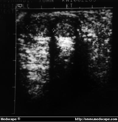
Figure 1. Ultrasonography of left hemiscrotum revealed 1.1 x 0-cm solid mass and second mass measuring 1 x 0.6cm. Both are well defined, ovoid, and with homogenous echogenicity consistent with either single bilobate testis or 2 separate testes.
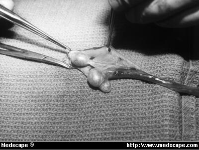
Figure 2. At surgery, 2 left testes evident within left hemiscrotum with thin septum of tissue connecting upper and lower epididymis.
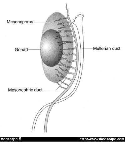

Figure 3. Normal embryology of developing testis, epididymis, and vas deferens from early, indifferent stage (A) to later, fully developed stage (B).
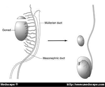
Figure 4. Leung type A polyorchidism.
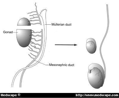
Figure 5. Leung type B polyorchidism.
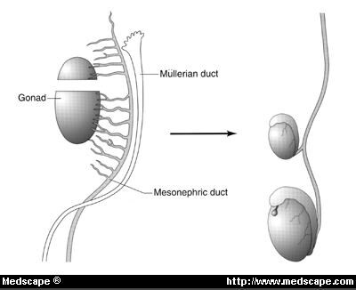
Figure 6. Leung type C polyorchidism.
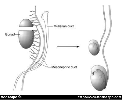
Figure 7. Leung type D polyorchidism.