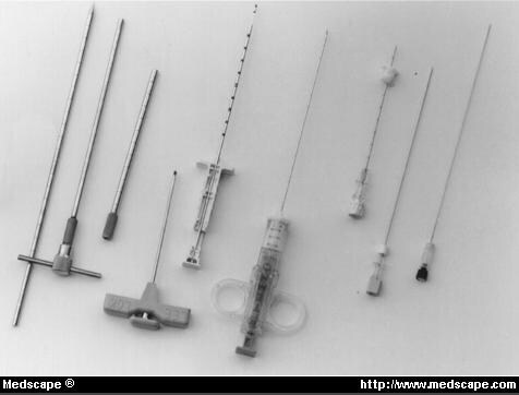
Figure 1. Instruments routinely used for closed needle biopsy (left to right): for bone, the Craig trephine set or Lee-lok Trephine (Hema Science Co., Minneapolis, Minn) are used. Core biopsy of soft tissue can be obtained with the Tru-Cut (Baxter Healthcare Co., Deerfield, Ill), or with the Adjustable Coaxial Temno (Bauer Medical, Inc., Clearwater, Fla). Fine needle aspiration is performed with the 18-gauge Crown (Medi-Tech, Inc., Watertown, Mass) or the 22-gauge Westcott (Becton Dickinson Co., Rutherford, NJ).
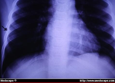
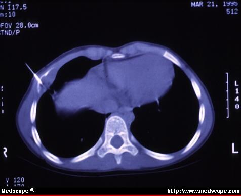
Figure 2. Five-year-old boy with painful destructive rib lesion. (A) Plain radiograph demonstrates expansion and destruction in right fifth rib. (B) Under general anesthesia, CT guided FNA confirmed eosinophilic granuloma, and lesion was injected with methylprednisolone.
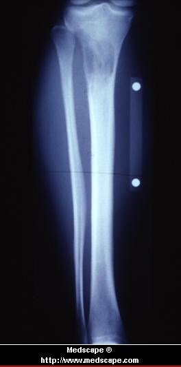
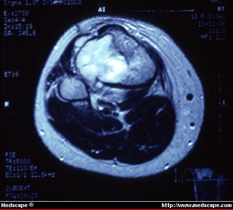
Figure 3. Eighteen-year-old woman with destructive proximal tibial lesion with associated soft-tissue mass, suspicious for osteosarcoma versus Ewing's. (A) Plain radiograph shows destructive metaphyseal tumor. (B) MRI demonstrates extra osseous soft-tissue mass suitable for core needle biopsy. Under general anesthesia core needle biopsy was performed, and frozen section demonstrated osteosarcoma. A Hickman catheter was then placed under the same anesthetic.
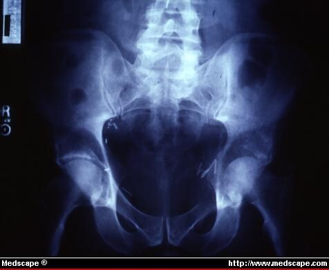
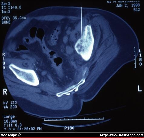
Figure 4. Sixty-year-old man with known prostatic cancer and pathologic fracture through left acetabulum. (A) Plain radiograph demonstrates destructive, poorly demarcated lesion in left acetabulum/superior ramus through which patient has sustained pathologic fracture. (B) Lesion was isolated and lytic. CT guided trephine biopsy was used to confirm prostatic metastases.
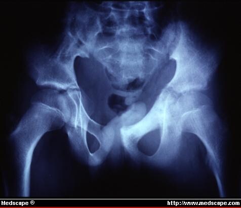
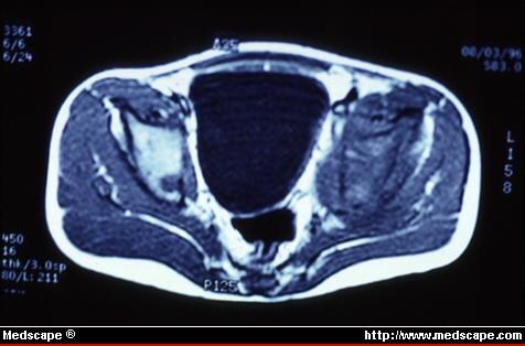
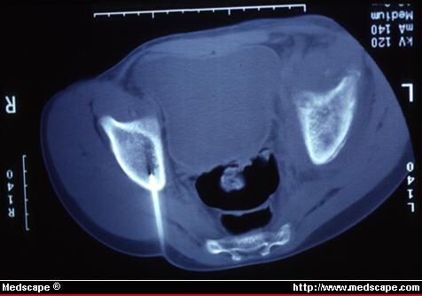
Figure 5. Thirteen-year-old boy with biopsy proven Ewing's sarcoma in left pelvis, underwent CT guided biopsy of suspected bony metastasis to right acetabulum. (A) Plain radiograph demonstrates primary left acetabular site. (B) MRI shows primary left site and right acetabular dome lesion. (C) Trephine-type needle biopsy of dome lesion confirmed bony metastasis.
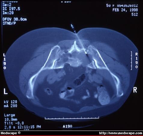
Figure 6. Fifty-year-old woman with minimally painful sacral lesion and no neurologic deficit. CT guided biopsy with a core needle. Note midline approach, in case sacral resection is later required. Pathology demonstrated benign neurofibroma.
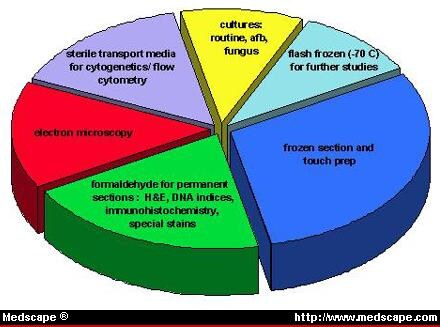
Figure 7. Allotment of core needle specimen. Frozen section, permanent section, and cultures are performed routinely. Further studies are based on the frozen section as deemed appropriate by the pathologist.