Figures for:
Current Management of Community-Acquired Pneumonia in Children: An Algorithmic Guideline Recommendation
[Infect Med 16(1):46-54, 1999. © 1999
Cliggott Publishing, Division of SCP Communications
]

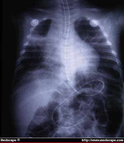
Figure 1. Hyperexpansion, parahilar peribronchial infiltrates, atelectasis, and hilar adenopathy in 6-week-old infant who rapidly progressed to respiratory failure. Tracheal aspirate was positive for RSV antigen by ELISA methodology.

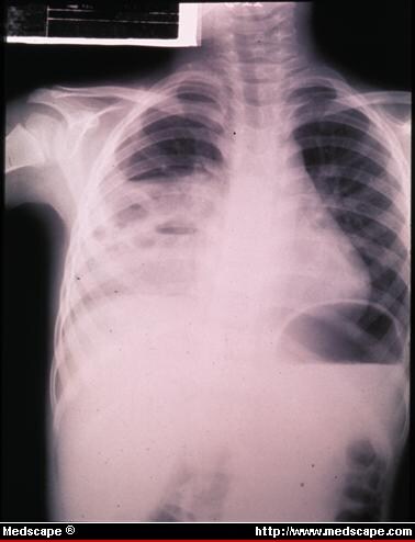
Figure 2. Consolidated right upper and middle lobe pneumonia with pneumatocele formation, most compatible with Staphylococcus aureus as etiology. Organism was recovered by needle aspiration of lung. This young infant subsequently developed large pleural effusion.

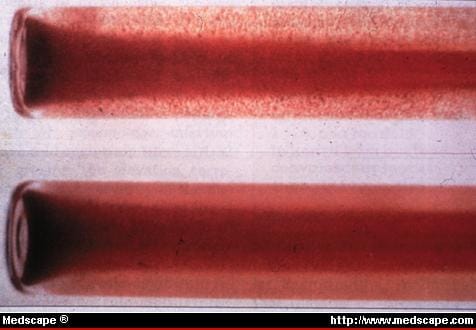
Figure 3. Rapid screening test for cold agglutinins: 4-5 drops of blood are collected in 60 × 7mm Wasserman tube containing approximately 0.2mL of NaEDTA. Tube is capped and placed in ice water bath for 30-60 seconds, after which it is tilted so that blood runs down wall of tube. Definite floccular agglutination seen with unaided eye (upper panel), which disappears upon warming to 37[ring]C (98.6°F) (bottom panel), is interpreted as positive. Control sample is useful for interpretation.

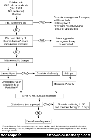
Figure 4. Inpatient and outpatient management of children with CAP.


